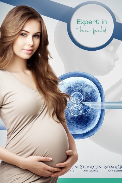
Treatments
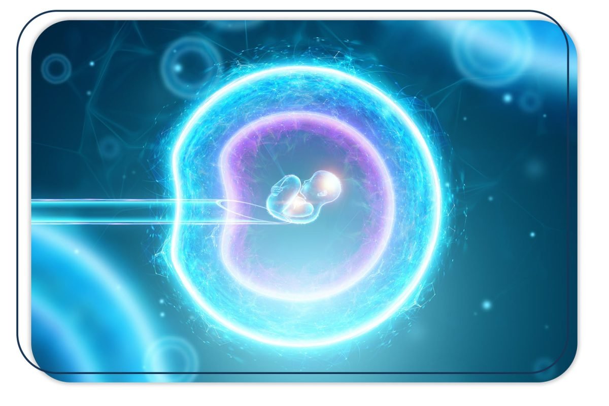
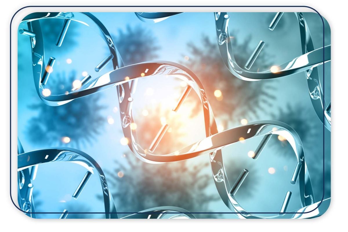
Mitochondrial Transfer( Mitochondrial Donation)
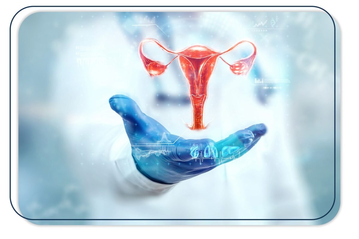
Assisted reproductive treatments can be performed to help health pregnancy. In case of low ovarian reserve, PCOS syndromes, many IVF failure without receiving any blastocysts or reason of mitochondrial mutation may cause infertility problem. Especially for advanced maternal age together with low ovarian reserve, oocyte( egg) quality and mitochondrial quality can decrease dramatically. Mitochondria is the energy supplier of oocyte and when oocyte has lack of energy embryo development will not process properly. Also, if there is mitochondrial mutation after the birth or genetical testing for many families need an alternative to egg donation to carry up their DNA for future generations. Mitochondrial transfer will create and alternative to egg donation to achiving healthy babies.
Mitochondria are essential organelles, with copies found in varying numbers in most cells in the body. Their most significant role is in serving as the cell’s primary energy source. Mitochondria contain their own DNA, consisting of 13 genes. These genes are involved in producing and transmitting cellular energy, and are crucial in the reproductive process. It’s well known that mitochondria become dysfunctional with advancing age, which directly affects embryonic development. Therefore, the transfer of genetic material from an oocyte or zygote that contains older mitochondria, to a younger one with more active mitochondria, can help restore normal embryonic development.
Mitochondrial transfer is highly specialized medical technology that required experience and special technology and equipment. The technology can offer help to families who affected from genetic diseases by mitochondrial mutation and also patients with advanced maternal age with poor egg quality and blastocyst outcome.
Mitochondrial Diseases and Mutations
PGD has been used in different ways, particularly to determine the amount of mitochondria that may carry the mutation as the fetus grows. This technique aims to transfer embryos with a low risk of mutant mtDNA on a 6-10 cell basis in trophectoderm biopsy samples obtained from embryos obtained using the IVF technique. However, PGD technology is limited in its ability to provide results only for mitochondrial mutations and diseases caused by nuclear DNA. Mutations in mitochondrial DNA increase the risk of affecting embryos due to maternal involvement of mitochondria. Due to the lack of techniques particularly for estimating the degree of heteroplasmy in mtDNA, it is not easy to predict the risks associated with mtDNA. In this regard, mitochondrial transfer techniques have been successful in ensuring a healthy subsequent live birth in generations where mtDNA mutations have been detected.
Aging Oocyte
Mitochondria have a crucial role in the process of aging and metabolic decline, especially as a major source of reactive oxygen species. Increased levels of reactive oxygen species result in less mitochondrial energy being metabolically available. As age progresses, molecular senescence progresses to an advanced stage as increased levels of ROS affect mtDNA genome proliferation and both the morphological capacity and metabolic capacity of the oocyte are reduced.(6) The increased level of aneuploidy, in particular, can be explained by the fact that, as meiosis is reactivated, a greater amount of intracellular mitochondrial energy will be needed, and this ATP energy is lower in older patients.
Advanced Maternal Age and Mitochondria
Recent changes in living conditions, changes in women’s social status, and postponement of childbearing due to career and occupational priorities have created the problem of achieving pregnancy at advanced reproductive age. The recognition that mitochondria are the center of quality power, especially in the oocyte cell, has confirmed that mitochondria play an important role in the problem of embryonic development at advanced age. In the normal process, paternal mitochondria are captured by autophagosomes and transferred to lysosomes for degradation after fertilization. Here, due to aging, mitochondria begin to show morphological changes and impaired activity. The loss of mitochondrial energetic activity at an older age has been associated with problems in the transition of embryos from cleavage to blastocyst at the cleavage stage of embryo development when energy requirements are high. The use of young mitochondria in these patients has been shown to significantly increase the rate of blastocysts obtained from embryos. In oocyte donation, it is observed that embryos obtained from oocytes obtained from young donors can easily achieve pregnancy when transferred to elderly patients. The most significant finding here is the high rate of euploid embryos in young patients, which leads to a higher chance of pregnancy success. Additionally, it has been observed that young oocytes had a high euploid rate, and euploid embryos were obtained from euploid oocytes. In both mouse and other animal experiments, it has been found that older oocytes had both a very low euploid rate and very low mitochondrial activity. It has also been noted that older women have much lower mtDNA at the zygote stage than younger women and that embryos from these zygotes do not reach the cleavage stage to the blastocyst stage.
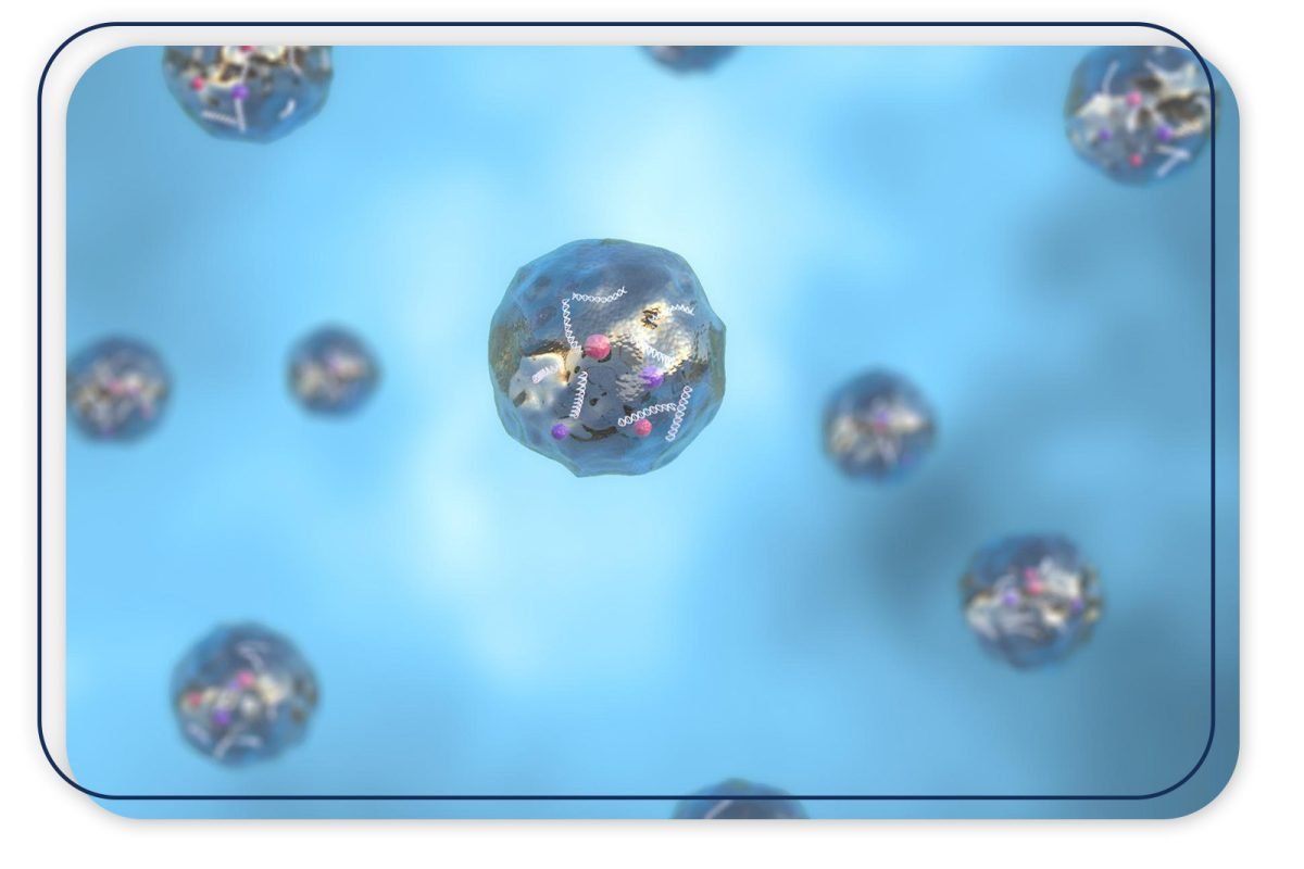
Mitochondrial Donation
Using a fertility-proven donor oocyte with healthy mitochondria, especially in diseases related to mitochondrial mutations, eliminates the risks and ensures the formation of healthy embryos. For various reasons, many patients with mitochondrial disease are diagnosed late, and due to the advanced age at which treatment is started, both the quality and quantity of oocytes are limited. Naturally, blastocyst development is very limited in patients with this advanced age characteristic. With the identification of good embryo development in these elderly patients, it was realized that mitochondrial transfer may be an alternative to oocyte donation in elderly patients. This method utilizes the mitochondrial potential of high-quality donor oocytes to produce high ATP energy, resulting in high-quality blastocysts in many patients. However, progress in correcting chromosomal defects in embryos obtained by this method is limited.
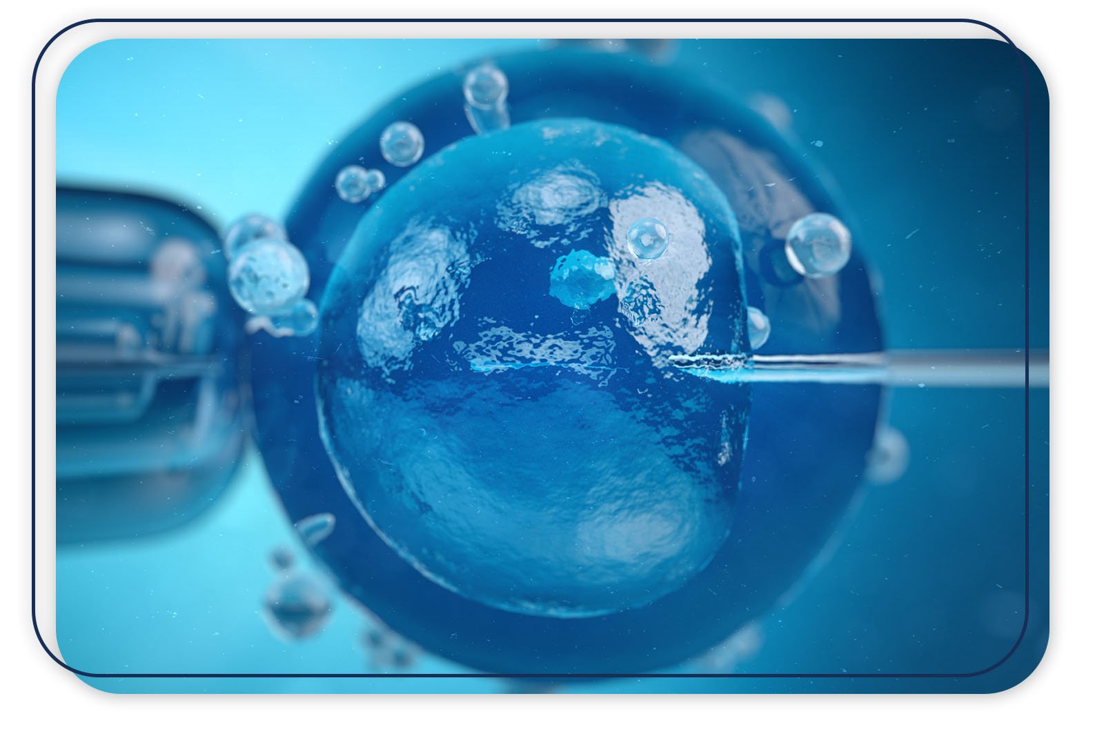
Meiotic Spindle Transfer
The meiotic spindle is the structure that forms during anaphase between meiosis 1 and meiosis 2, and it also serves to transport centrosome proteins. The meiotic spindle has a fundamental role in the development and genetic structure of the oocyte. The formation of the meiotic spindle by the separation of the chromosome package in the first anaphase phase indicates that the transfer of chromosomal structure occurs in the later stages of embryonic development. Therefore, it can be hypothesized that the transfer of the meiotic spindle from the oocytes of older women to the young donor oocyte results in a much better morpho-kinetic development with higher mitochondrial energy and even a lower aneuploidy rate.
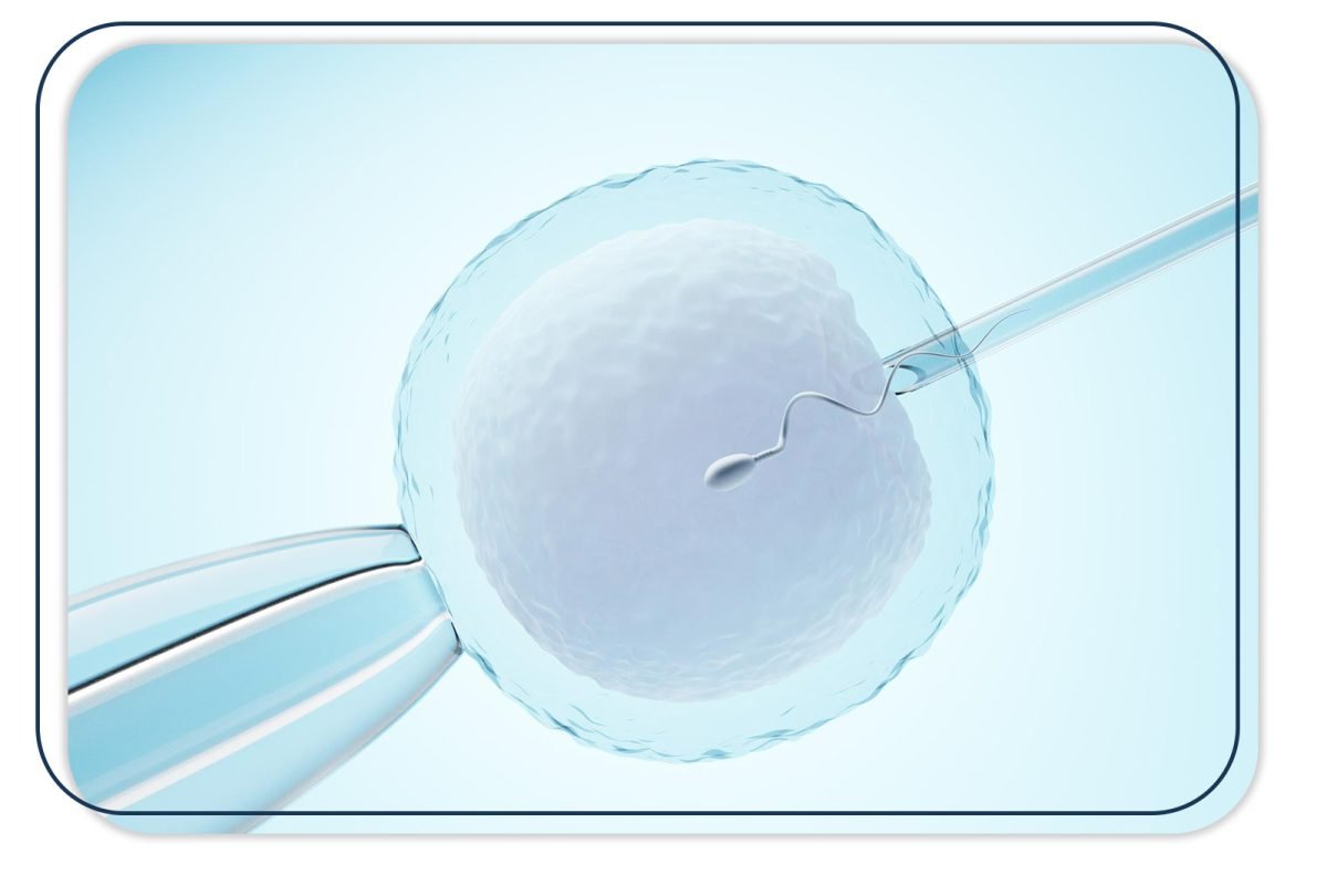
Oocytes from an older patient and young donor stimulated with the same trigger timing should be prepared for transfer by determining the location and size of the spindle using spindle imaging software and polarizer, if the oocyte is mature, immediately after separation of the cumulus complex structure. The 24-hour prior addition of Cytochalasin B to the incubated single-step culture medium protects the zona permeability and cytoplasmic structure of the oocyte during micromanipulation. The spindle is removed using spindle view technology with the help of a small transitional zone to be opened using the laser hatching method in the zone of the oocytes taken from both the elderly patient and the oocyte donor, and the spindle is removed and subjected to fusion in the HVJ-E virus. This process aims to eliminate DNA fragments from the cytoplasmic structure around the spindle and to transfer the spindle into the donor oocyte as a compact DNA carrier. Instead of the spindle removed from the donor oocyte, the spindle structure of the older patient is transferred similarly, and the oocyte is incubated in the prepared solution for a while to ensure the formation of a complete karyoplast. After this procedure, the oocytes are incubated for 1 hour, and then fertilization is performed using the piezzo ICSI procedure.
Pronuclear Transfer
In patients of advanced maternal age, pronuclear transfer can be used as an alternative to spindle transfer, especially in the absence of a history of fertilization issues. The pronuclear transfer would be a good alternative, especially when oocyte quality is poor, and meiotic spindle structure cannot be observed. Another advantage of the pronuclear transfer procedure is the possibility of working on frozen zygote samples. Here, the zygote of an elderly patient can be used frozen, whereas a fresh zygote with fresh donor oocyte collection and a fresh fertilization process needs to be preferred for micromanipulation. Particularly after the ICSI procedure, early pronucleus detection ensures a better quality and successful micromanipulation procedure. Nocodozale and Cytothalasine B are added to the single-step culture medium incubated 24 hours before the procedure to preserve the zona permeability and cytoplasmic structures of donor and elderly patient zygotes in the petri dish. A crossing point is created using laser hatching on the zona of the zygotes, and with the help of a biopsy pipette, nuclei are extracted from both zygotes and introduced into the HVJ-E virus for fusion. The main purpose of this procedure is to transfer the compact DNA structure from the nucleus into the donor zygote by neutralizing the cytoplasmic fragments around the nuclei. After the transfer, the zygotes are left for a certain period to complete the karyoplast process and then transferred into the single-step culture medium, and embryo development is monitored.
Contact Us
Details available with Every Demo
Projektet e ndërlidhura
- Stem Cell Treatments

- Stem Cell Treatments
- Stem Cell Treatments
- Stem Cell Treatments
- Stem Cell Treatments

- Stem Cell Treatments

